Contact us
Address
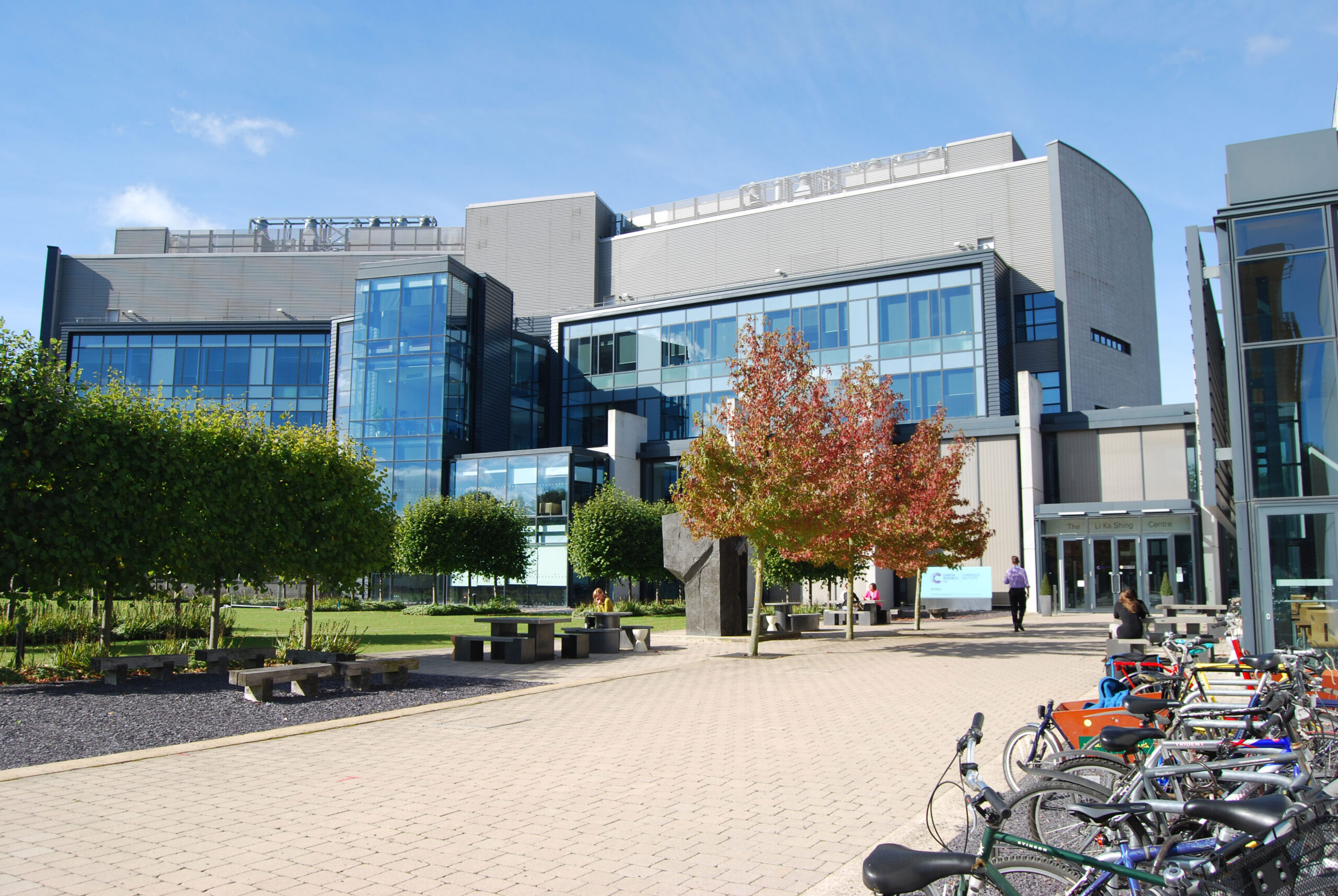
Visit us at CRUK Cambridge Institute
University of Cambridge, Li Ka Shing Centre, Robinson Way, Cambridge CB2 0RE

Visit us at CRUK Cambridge Institute
University of Cambridge, Li Ka Shing Centre, Robinson Way, Cambridge CB2 0RE
Designed for data-intensive research, Darkroom combines interactive analysis environments, high-performance computing resources, workflow automation, and secure data access within a single integrated platform.
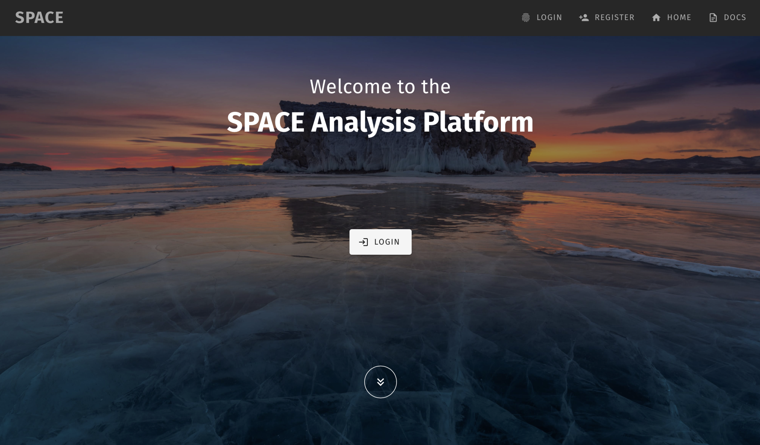
Modern scientific projects generate terabytes of data from imaging systems, sequencing instruments, telescopes, and large-scale experiments. Analysing these datasets requires significant computing power and specialised software environments.
The SPACE Analysis Platform removes these barriers by providing:
To complement the cloud platform, the SPACE Desktop Client provides a secure gateway for data ingestion and management. It facilitates the seamless upload of large-scale, multi-modal microscopy images directly to the cloud while automatically creating and tracking detailed database entries.
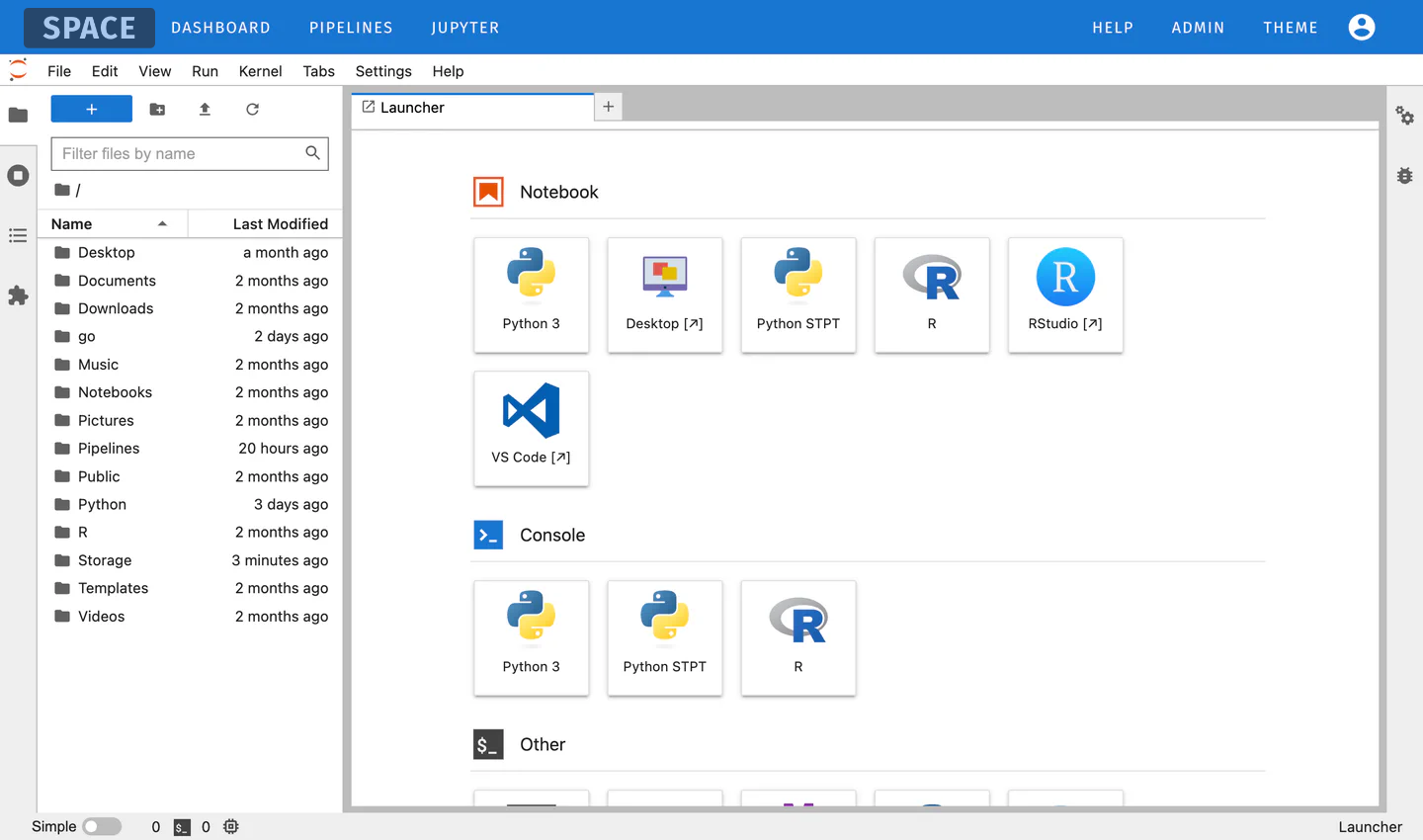
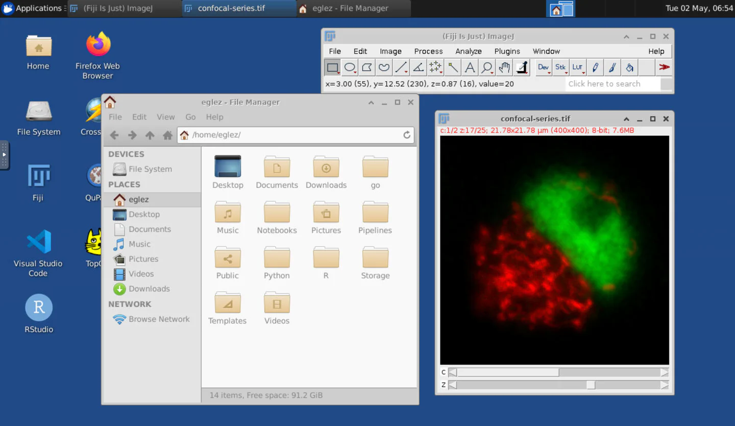
Launch fully managed analysis environments directly from your browser.
Supported environments include:
No local installation required. Compute resources are provisioned on demand.
From exploratory analysis to large-scale production workloads, SPACE provides access to scalable compute infrastructure.
Capabilities include:
SPACE provides integrated access to large research datasets through modern object storage technologies.
Features include:
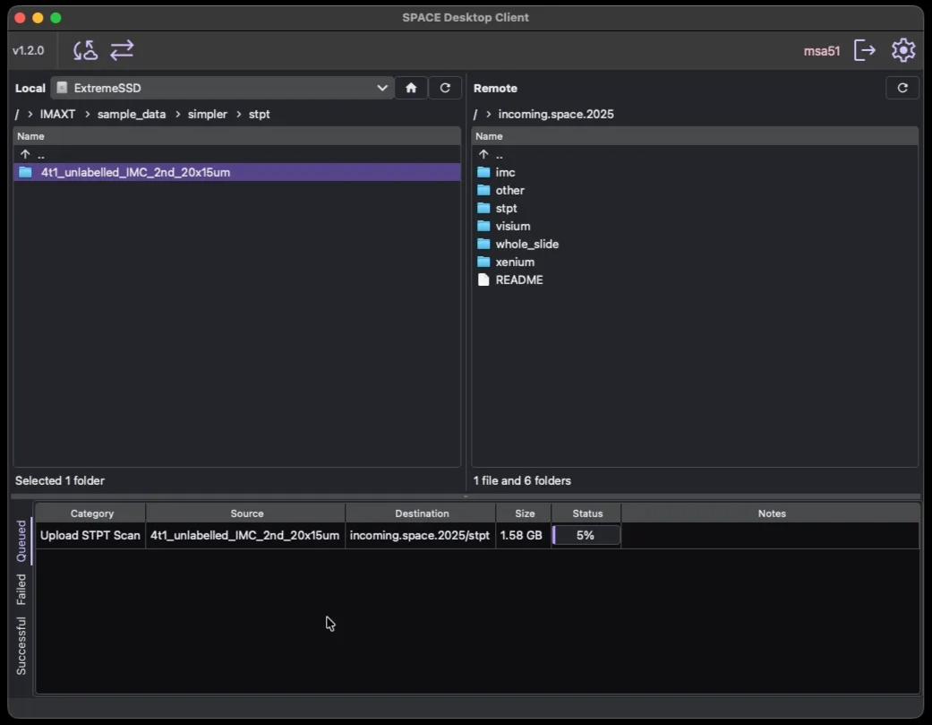
To complement the cloud platform, the SPACE Desktop Client provides a secure gateway for data ingestion and management. It facilitates the seamless upload of large-scale, multi-modal microscopy images directly to the cloud while automatically creating and tracking detailed database entries.
Opal Studio
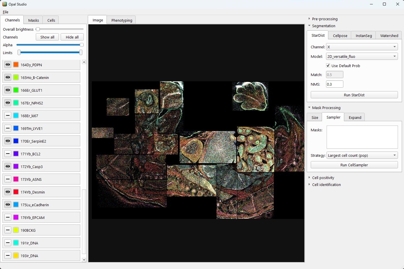
Opal Studio is a cross-platform image visualisation and analysis application for highly multiplexed imaging data, including Imaging Mass Cytometry (IMC), with support for OME-TIFF and SpatialData/Zarr formats. It provides responsive multi-channel rendering for large images and brings together common spatial biology workflows in a single interface, including channel preprocessing, cell segmentation, AI-based marker positivity detection, phenotype gating, and clustering.
Opal Studio also integrates several related tools developed by our group. These include a converter which generates virtual bright-field images from multiplexed images, and a consensus cell mask merging tool for combining outputs from multiple segmentation algorithms into a single robust cell segmentation mask.
Cyto Studio
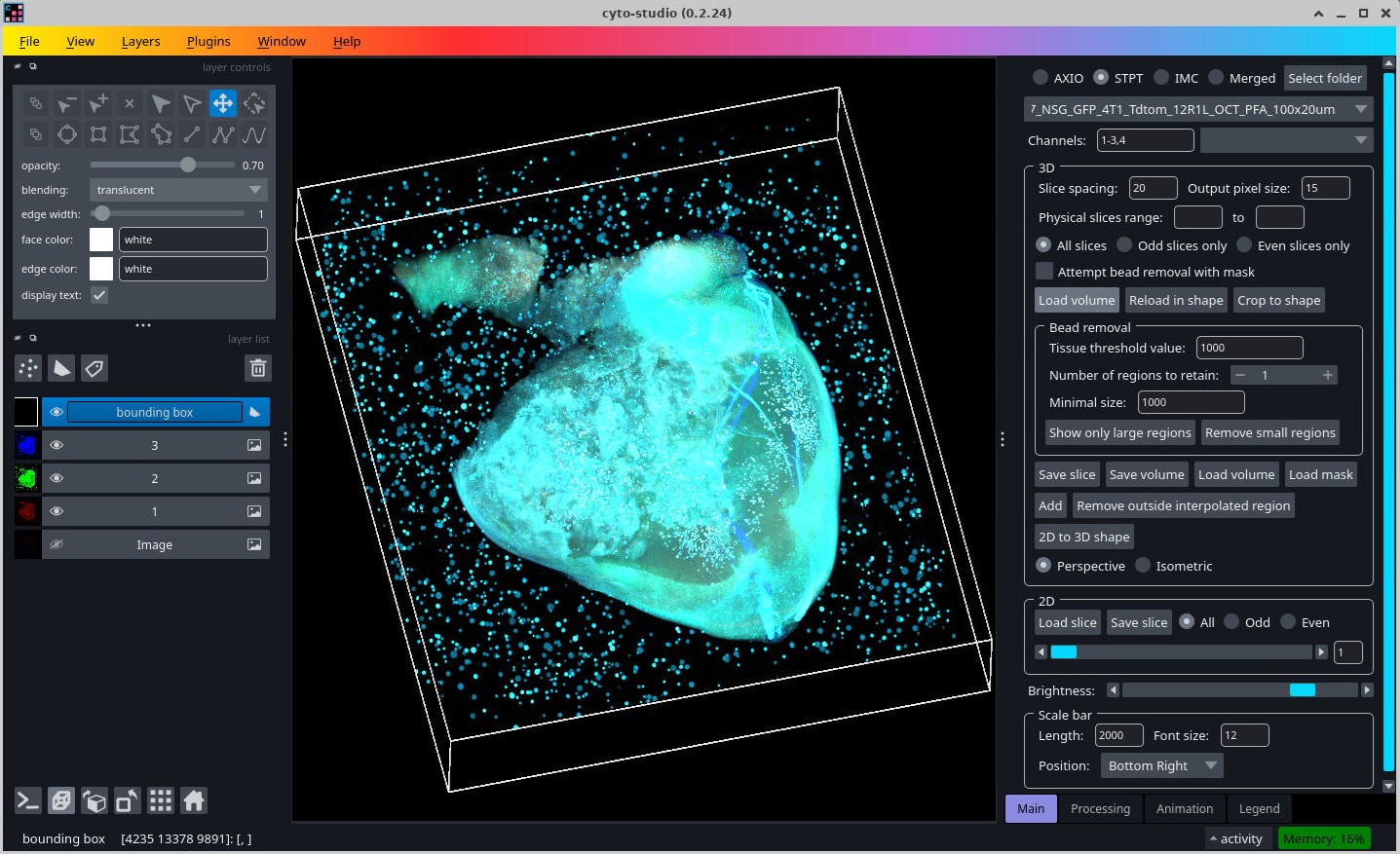
Cyto Studio is a Napari-based viewer for exploring large 2D and 3D multiplexed image datasets. It was developed to make image browsing more accessible by providing an interactive interface for selecting datasets, loading image channels, and inspecting images without requiring users to write custom visualisation code. The software runs on the same system where the imaging data is stored and can be accessed through a virtual desktop environment, thereby reducing data-transfer overhead and optimising image loading times.
References
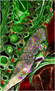
Through partnership with several teams across the CI, SPACE has validated over 300 antibodies for IMC use in FFPE tissue as well as several for fresh frozen and agarose embedded tissues. Although antibody conjugation to metal isotopes yields the best results with carrier-free antibodies, we have had many successful validations of antibodies that contain BSA, sodium-azide, and glycerol. The process for validating a new antibody for IMC includes IHC staining verification to confirm expected antibody binding pattern, conjugation to a metal isotope, and evaluation of staining at varying dilutions and across a range of tissue types. Where possible, SPACE has utilised TMAs for many IMC antibody validations. Our current TMA contains over 20 different human tissue types, including both tumour and normal tissue.
Dr. Alireza Molaeinezhad – Application Specialist
Alireza Molaeinezhad is an Assistant Research Professor at the University of Cambridge whose work combines astrophysics, AI, image analysis, and large-scale scientific computing. His interdisciplinary research applies advanced image processing, computer vision, and machine learning techniques to challenging problems across both astrophysics and biomedical sciences. He develops computational methods and scalable data analysis systems for multimodal imaging, complex scientific datasets, and high-performance research infrastructures.
Dr. Tristan Whitmarsh – Application Specialist
Tristan Whitmarsh is an interdisciplinary researcher working at the intersection of artificial intelligence and biomedical imaging, with more than a decade of experience in biomedical image analysis. He is currently a Research Associate at the Institute of Astronomy, University of Cambridge, where he works on the Cancer Grand Challenges IMAXT project and its extension through the Spatial Profiling and Annotation Centre of Excellence (SPACE). His research focuses on developing image analysis and deep learning methods for high-resolution, high-dimensional imaging, including multiplexed immunofluorescence, imaging mass cytometry, serial two-photon tomography, as well as traditional brightfield imaging. He develops computational approaches for cell and tissue segmentation and spatial analysis to better understand the tumour microenvironment. He also develops and maintains software tools that help researchers visualise, annotate, and interpret complex spatial imaging data, including Cyto Studio and Opal.
Dr. Melis Irfan – Application Specialist
Melis Irfan is physicist who specialises in image and signal processing. She is a research associate at the Institute of Astronomy, University of Cambridge working alongside researchers at the Precision Breast Cancer Institute and CRUK Cambridge Institute. Her work in cosmology focuses on the formation of Large-Scale Structure in the Universe and within cancer research she uses computer vision techniques to aid cancer diagnosis, treatment, and research.
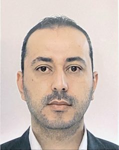
Dr. Mohammad Al Sa’d – Software Developer
I am a Medical Software Developer focused on creating user-centric applications and essential data wrangling tools for the SPACE project. By combining iterative user feedback with technical backend refinement, I build software that simplifies data management and enhances the overall user experience. My work ensures that large-scale datasets remain accessible and adaptable, covering the full development lifecycle from API design and desktop client expansion to facilitating seamless data acquisition and cross-format interoperability.
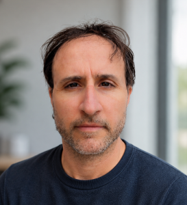
Dr. Eduardo Gonzalez-Solares – Senior System Administrator and Developer
Eduardo is an Assistant Research Professor at the University of Cambridge with a background in Astronomy and Data Intensive Science. He has extensive experience in large-scale data processing and analysis, machine learning, and data management. His work combines scientific research with software engineering, leading the development of analytical tools and the frontend and backend systems that power the SPACE analysis platform.
https://orcid.org/0000-0001-9123-0626
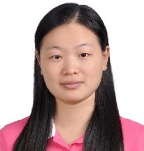
Dr. Qiuchen Meng – Bioinformatician
Qiuchen Meng is a bioinformatician working at the intersection of artificial intelligence, spatial omics, and biomedical data analysis. She is currently a postdoctoral researcher at the University of Cambridge and CRUK Cambridge Institute. Her work focuses on imaging mass cytometry, spatial proteomics, single-cell multi-omics, and AI-assisted analysis systems, with applications in tumour microenvironment research. She has developed complete IMC analysis pipelines from raw imaging data to quality control, segmentation, spatial phenotyping, and downstream analysis. Her current work aims to build reproducible and AI-assisted computational frameworks that make complex spatial omics analysis more accessible, reliable, and interpretable.
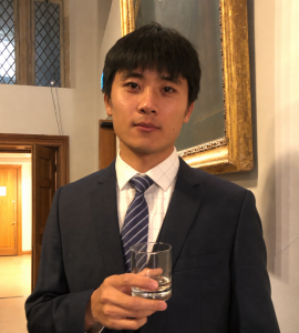
Dr. Kui Hua – Senior Bioinformatician
Kui Hua is an Assistant Research Professor at the Cancer Research UK Cambridge Institute (CRUK-CI) and Chief Bioinformatician of the Spatial Profiling and Annotation Centre of Excellence (SPACE-lab). He earned his Ph.D. in the Department of Automation at Tsinghua University in 2021, followed by postdoctoral training with Dr John Marioni and Professor Greg Hannon at CRUK-CI and EMBL-EBI. Kui’s research sits at the interface of computational biology, spatial omics, statistical modelling, and artificial intelligence. He develops computational methods and platforms for the integrative and comparative analysis of cohort-level spatial multi-omic data, with a particular focus on understanding how cells coordinate to form tissues, organs, and complex organisms, and how these spatial relationships are altered during development, injury, disease, and cancer. His current work aims to move beyond descriptive analysis of individual samples towards scalable frameworks that can compare tissue organisation across patients, conditions, perturbations, and modalities. By combining spatial transcriptomics, proteomics, imaging, machine learning, and statistical modelling, his long-term goal is to uncover general principles of cellular organisation and communication, and to translate these insights into improved understanding of cancer biology and therapeutic response.
https://orcid.org/0000-0003-2228-7025
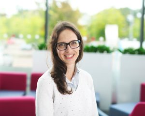
Dr. Giorgia Battistoni – Postdoctoral Research Associate
Giorgia is a postdoctoral fellow in the laboratory of late Prof. Greg Hannon at the University of Cambridge (UK), where she works at the intersection of epigenetics, technology development and spatial biology. Giorgia obtained a BSc and MSc in Molecular and Cell Biology from the University of Pisa and Scuola Normale Superiore di Pisa in 2013, and a PhD in Biological Sciences from CSHL School of Biological Sciences (Cold Spring Harbor, NY) in 2019. Her focus is to understand how regulatory programmes for gene expression are modulated by environmental cues — and whether such adaptations are transmitted across generations. Her work approaches this question from two complementary directions: I. deciphering the epigenetic control of cell fate in the mammalian embryo and pathological samples, and II. building the tools necessary to study it with the resolution and scale the question demands.
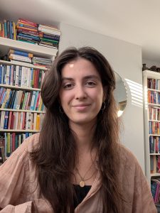
Mirta Vojnovic – PhD student, shared with Richard Mair laboratory (University of Cambridge)
Mirta Vojnovic is a PhD student at the University of Cambridge, working on the SPACE-Glioma project in collaboration with the Mair Lab. Her research applies spatial profiling technologies to clinical samples to investigate malignant transformation and tumour evolution in glioma. She previously completed her MPhil in Medical Sciences with the Mair Lab (University of Cambridge) and holds a First-Class BSc in Neuroscience from the University of Bristol. Her work integrates in vivo and in vitro models with spatial approaches to study tumour–microenvironment interactions and identify therapeutic vulnerabilities in neuro-oncology.
https://orcid.org/0009-0005-6422-5422
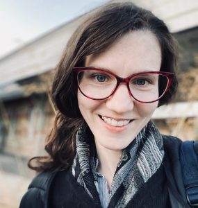
Dr. Oksana Sutyagina – Research Assistant
Oksana Sutyagina studied Cell Biology and Histology at the Faculty of Biology, Lomonosov Moscow State University, where she later completed a PhD in Cancer Biology in 2019. Her subsequent research focused on morpho-functional changes in astrocytes during ageing and glioma formation, and ( in parallel) investigating the cell type-specific role of the Hippo–YAP signalling pathway using organoid models. Her favourite technique is multicolour Flow Cytometry. In March 2026, she joined the SPACE team as a Research Assistant with a strong interest in gaining hands-on experience in spatial protein profiling and spatial transcriptomics technologies.
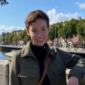
Phoebe Hicks – Research Assistant
Phoebe is a research assistant with a background in clinical histology and has a wide range of experience from molecular research to pharmaceutical QC. She completed her MSc in Biomedical Science at London Metropolitan University in 2020. Since 2024, she has been part of the SPACE team at CRUK and is responsible for many of the technical aspects of both internal and external projects including assisting with antibody conjugations to metal isotopes, cryosectioning, tissue staining, and IMC imaging.
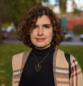
Atefeh Fatemi – Scientific Associate
Atefeh joined the Space/Formerly the IMAXT lab in 2020 and is currently a Scientific Associate. She manages the daily operations of Serial Two-Photon Tomography, including sample preparation, data processing, and data analysis. She earned an MSc in Cancer from UCL, where she focused on CAR-T cell therapy, and her interests are spatial cancer biology and cancer immunology
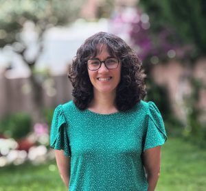
Dr. Marta Paez Ribes – Principal Scientific Associate
Marta Paez-Ribes studied biology in Barcelona, where she later obtained a PhD in biomedicine before moving to the University of Toronto as a postdoctoral fellow to investigate tumour angiogenesis. In 2015 she joined the University of Cambridge where she has been focused in unravelling the crosstalk between tumour cells and their microenvironment in the context of metastasis, with a special interest in spatial biology.
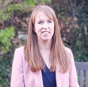
Dr. Claire Mulvey – Principal Scientific Associate
Claire Mulvey received her PhD in Pharmacology from University College Dublin, Ireland. She subsequently moved to London to pursue her interest in proteomics as a postdoctoral fellow in the Division of Medicine at University College London (UCL). Claire then joined the Cambridge Centre for Proteomics, Department of Biochemistry, University of Cambridge in 2011, where she continued her postdoctoral research in the field of mass-spectrometry-based subcellular proteomics. In 2018, she became an early member of the IMAXT Cancer Grand Challenge team at the CRUK Cambridge Institute. There, she optimised workflows for spatial profiling, including 3D volumetric imaging (serial two-photon tomography) and imaging mass cytometry (IMC), to generate molecularly annotated maps of tumours. Claire’s research interests focus on integrating state-of-the art spatial and imaging technologies to address key questions in cancer biology and support the goals of the SPACE projects.
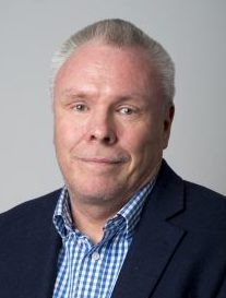
Prof. Nicholas Walton – SPACE co-lead investigator
Nicholas Walton is an astrophysicist at the Institute of Astronomy (IoA) with interests in galactic astrophysics and exoplanet systems. He leads the UK development teams producing the advanced data pipelines for major space and ground based facilities, including the photometric processing for the ESA Gaia mission, the exoplanet analysis systems for the ESA PLATO mission, and the team responsible for the analysis systems for the ESO MOONS and 4MOST facilities. He was a co-investigator of the IMAXT Cancer Grand Challenge project, and led the data team at the IoA.
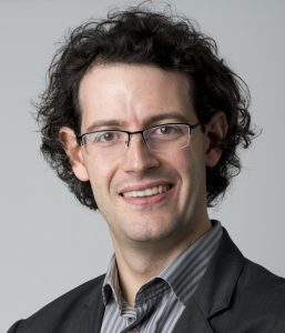
Dr. Dario Bressan – SPACE co-lead investigator
Dario Bressan started his career at the Scuola Normale Superiore of Pisa, Italy, where he graduated in Neurobiology. He then moved to Cold Spring Harbor Laboratory (NY), where he obtained a PhD in molecular biology working on the development of novel technologies for spatial profiling with Dr. Greg Hannon. He moved to the CRUK Cambridge Institute of the University of Cambridge in 2014, and he was a co-proposer of the IMAXT project, which was selected as one of the first four cancer grand challenge initiatives. From 2017 to 2024 he has been the head of the central hub laboratory for IMAXT, developing and optimising new technologies and applying them to breast cancer biology. In 2024 he spearheaded the transformation of IMAXT into the Spatial Profiling and Annotation Centre of Excellence, which is now democratising access to spatial biology to collaborative projects worldwide. His focus aress are on microscopy, genomics, and data analysis.
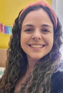
Dr. Carolina Mendonça – Programme Manager & Senior Scientific Associate
Carol is a molecular biophysicist with over a decade of experience in protein folding, dynamics, and interactions. She completed her MPhil and PhD at the University of Cambridge, followed by postdoctoral research at the University of California, Berkeley. In 2024, she joined Cancer Research UK Cambridge Institute as Senior Scientific Associate, before transitioning to a Programme Manager role within SPACE, where she supports all aspects of the group, its projects and collaborations, from setup to completion.